Fixed Anterior Alignment – Case Study: MK

Fixed Anterior Alignment by Dr Nick Simon

Dr Nick Simon is a former IAS teacher and Mentor. He was a delegate on the first cohort of the Advanced Fixed Course taught by Prof Ross Hobson, and this case won the Best Advanced Case at IAS Academy’s 2018 international symposium in London. This case shows the advantage of a systematic educational pathway and mentorship by an orthodontic specialist giving advice and confidence to the treating dentist—in this case, Prof. Ross Hobson from IAS Academy’s online mentoring forum.

CONTACT INFORMATION

The Ealing Dental Practice
6 Drayton Green Road,
West Ealing,
London,
W13 8RY

Website: Click Here

Case Summary

This is an early case on my Advanced Course journey.

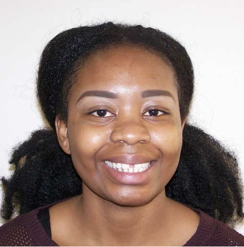
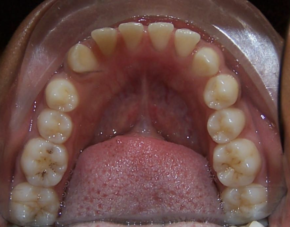
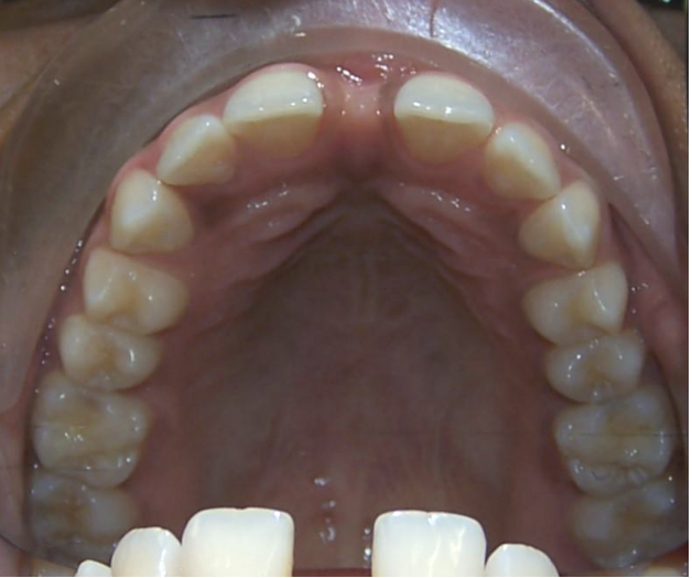
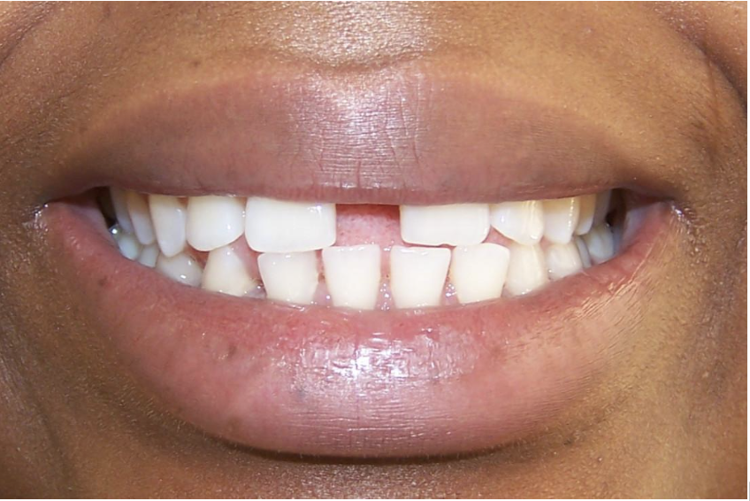
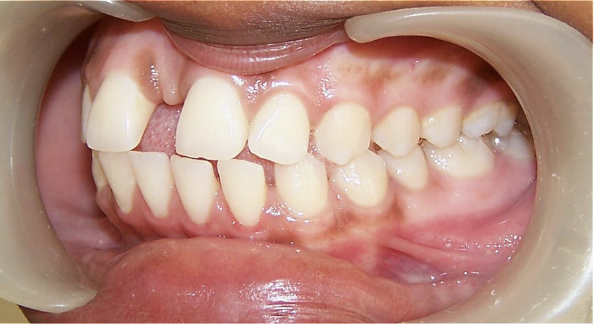
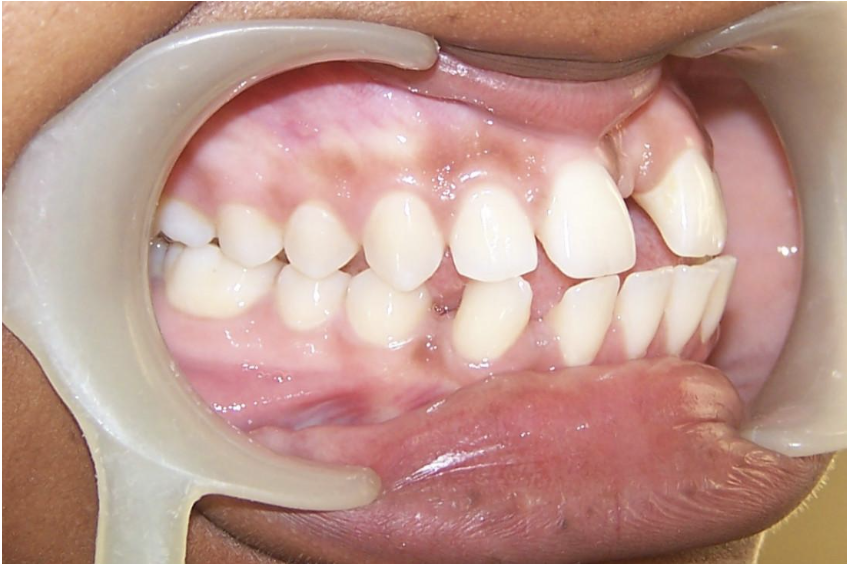
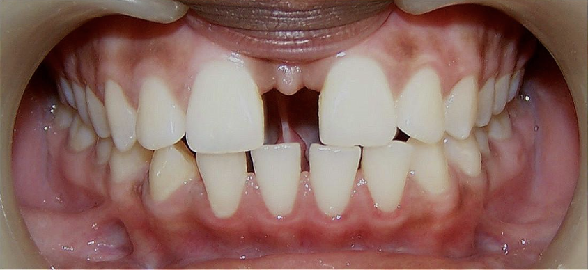
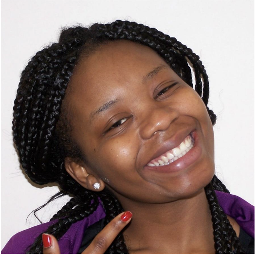
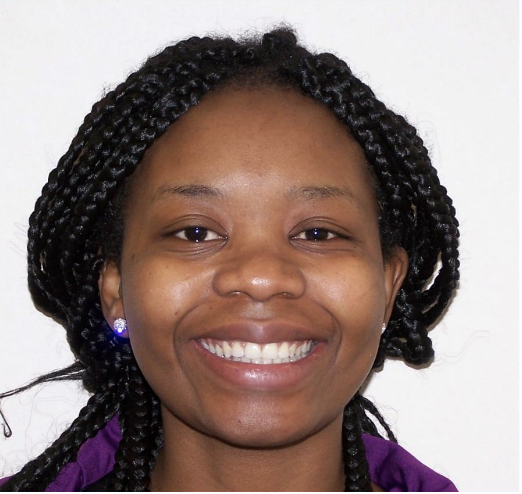
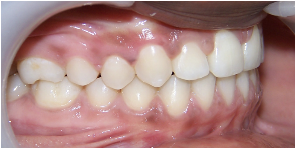
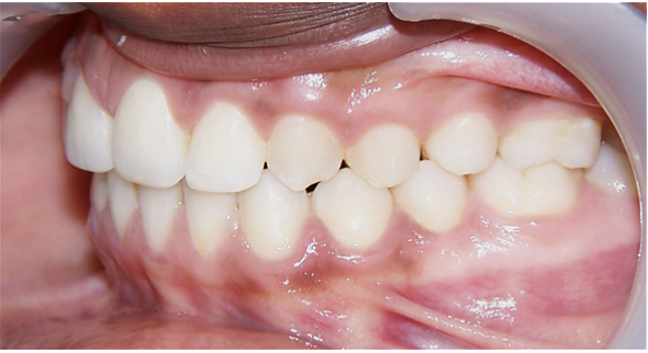
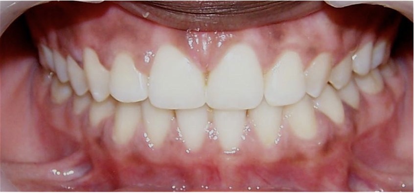
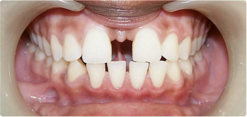
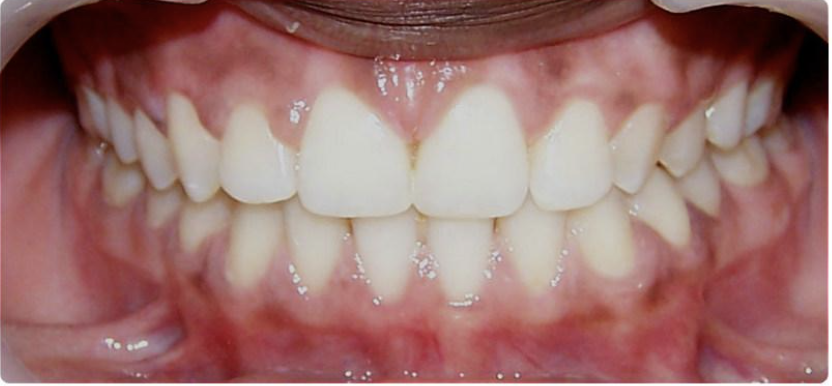
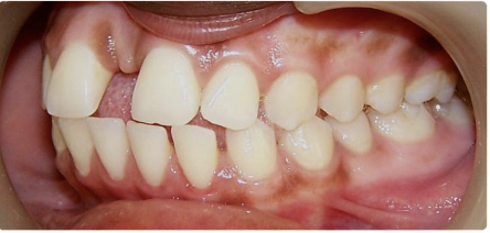
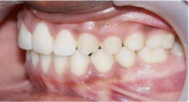
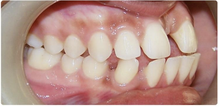
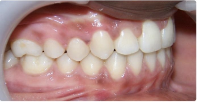
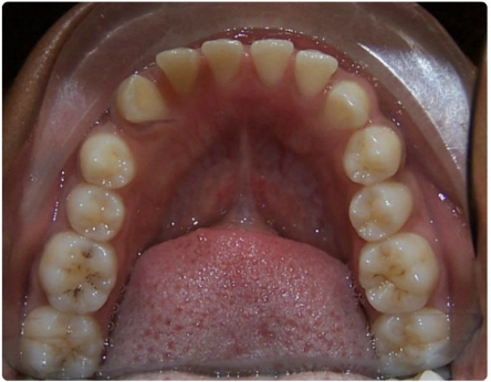
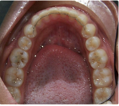
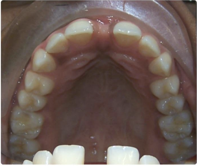
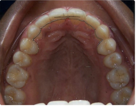
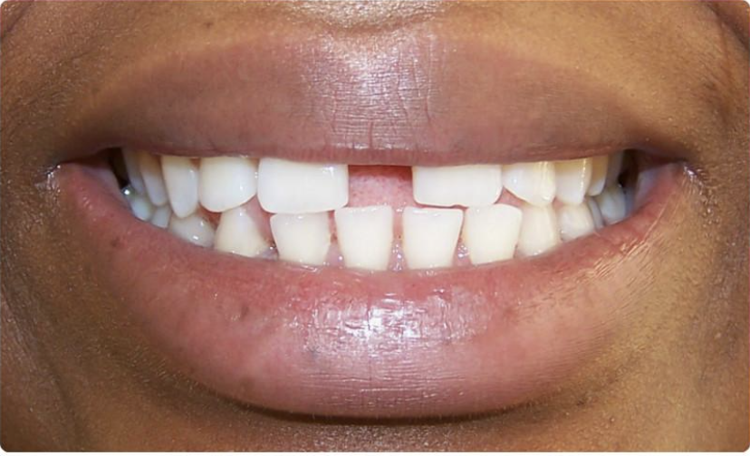
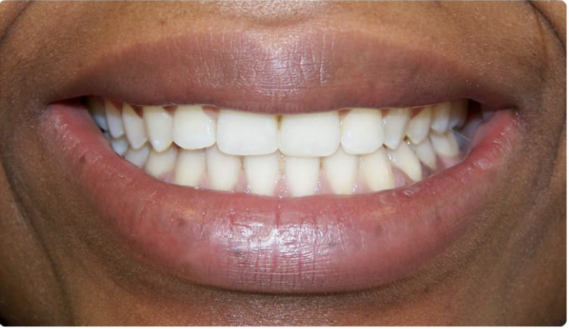
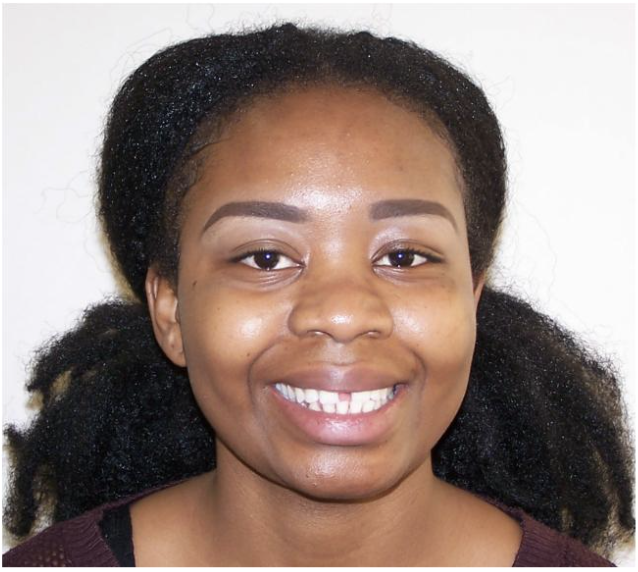
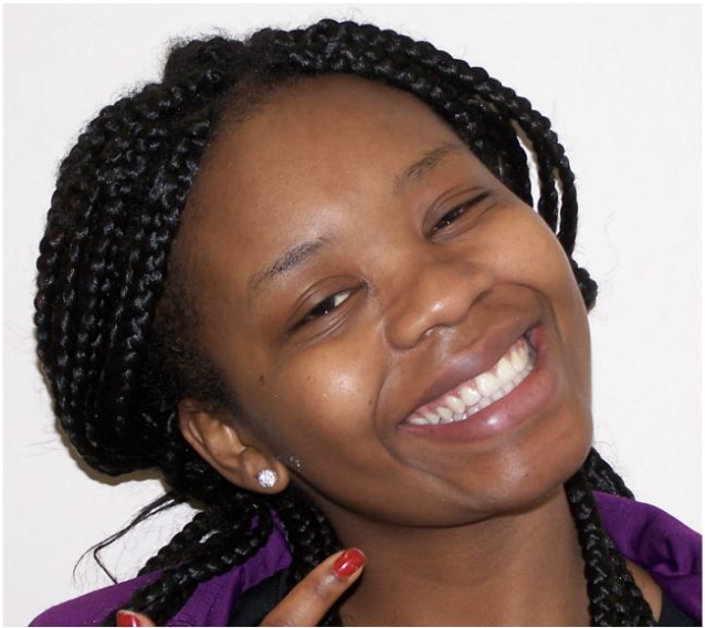
My patient was referred by her friend who had completed orthodontic treatment with myself. Her chief complaint were the gaps between her teeth which she had longed to have closed since her teenage years. The gaps were hereditary and with the tongue being a major soft tissue aetiological factor, retention would need to be planned carefully.

This case shows the advantage of a systematic educational pathway and mentorship by an orthodontic specialist giving advice and confidence to the treating dentist.

PRE-TREATMENT PHOTOGRAPHS: EXTRA-ORAL
PRE-TREATMENT PHOTOGRAPHS: INTRA-ORAL
Measurement Result
Skeletal Pattern Class I
FMPA Average
Lower Face Height Average
Facial Asymmetry No
Soft Tissues Details: Competent Lips, low smile line, acute NLA
Teeth Present All teeth present
Incisor Classification Class III
Overjet 1mm
Overbite 0%
Centrelines Upper and lower left by 1.5mm
Displacement on closure No
Molar Relationship Right Class I, Left Class I
Canine Relationship Right Class I, Left Class I
Crossbite No
Spacing Upper Arch 8mm, Lower Arch 10mm

Problem List

Spacing

Class 3 Incisors

Reduced overjet

Reduced overbite

Proclined incisors

Low lip line

Treatment

Fixed appliance therapy. Using PURITY® SINGLE CRYSTAL SAPPHIRE BRACKETS Roth .022.
Mentorship was provided on the Advanced Fixed Brace forum by Prof Ross Hobson.

Key Stages In Treatment Progress

Date Stage
June 2016 Bond up
Aug 2016 19 x 25 Niti, Start powerchain
Feb 2017 Upper Powerchain on hold, start Cl3 elastics
Oct 2017 Spaces Closed, detailing
March 2017 Debond / Fixed Retainers Placed

Mid Treatment Photographs

Bond up.
Laceback 1st Molar to Canine in each quadrant. 016 Niti in both arches.

MID-TREATMENT PHOTOGRAPHS: INTRA-ORAL

Below: Concern re lack of overjet. Uppers on hold and laceback  used to maintain position. Lower archwire hooks for Cl 3 elastics. Aim to keep lower space closure ahead of uppers.

MID-TREATMENT PHOTOGRAPHS: INTRA-ORAL

Below: Detailing required for all upper incisors. The tip is off.

MID-TREATMENT PHOTOGRAPHS: INTRA-ORAL
POST-TREATMENT PHOTOGRAPHS: EXTRA-ORAL
POST-TREATMENT PHOTOGRAPHS: INTRA-ORAL

Critical Appraisal

Assessment

I did not have my patient turn fully for the lateral profile photo. This shows strain on the neck muscles and I since learnt to position the patient more favourably. The patient had a higher lip line than the pre treatment photos depict. Preop lateral retracted photos were also not properly orientated.

Treatment

The patient was very compliant and progression was smooth.The patient was warned prior to treatment that extractions may be indicated should she slip into reverse overjet. This did not occur, yet Cl 3 elastics helped to maintain positive overjet.

I am pleased that the Class 1 canine and molar relationships were maintained. The initial photos may not have shown the Class 1 relationship.
A specialist from a previous ortho course recommended double fixed retainers in a large diastema case, and this drew some discussion on the Forum.
I have recently reviewed the patient and stability has been maintained.

The tip of the upper incisors could have been marginally improved. I had spent some time improving them in the detailing by resiting brackets. I need to learn how to fine tune with wire bending.

Comparison of before and after photos.

PRE-TREATMENT PHOTOGRAPHS: INTRA-ORAL
POST-TREATMENT PHOTOGRAPHS: INTRA-ORAL
PRE-TREATMENT PHOTOGRAPHS: INTRA-ORAL
POST-TREATMENT PHOTOGRAPHS: INTRA-ORAL
PRE-TREATMENT PHOTOGRAPHS: INTRA-ORAL
POST-TREATMENT PHOTOGRAPHS: INTRA-ORAL
PRE-TREATMENT PHOTOGRAPHS: INTRA-ORAL
POST-TREATMENT PHOTOGRAPHS: INTRA-ORAL
PRE-TREATMENT PHOTOGRAPHS: INTRA-ORAL
POST-TREATMENT PHOTOGRAPHS: INTRA-ORAL
PRE-TREATMENT PHOTOGRAPHS: INTRA-ORAL
POST-TREATMENT PHOTOGRAPHS: INTRA-ORAL
PRE-TREATMENT PHOTOGRAPHS: EXTRA-ORAL
POST-TREATMENT PHOTOGRAPHS: EXTRA-ORAL

Conclusion

The IAS advanced course was the best course I have completed. It gave me the ability to treat a whole range of orthodontic cases that previously I would have referred. This case is just one example of that. I would encourage anyone who has had some experience with fixed braces to join this pathway, you will not regret it. The mentoring is excellent and you will feel confident that you have the support and advice both during and after the course. That is so important to us GDP’s..to make us feel safe, plus you’ll  make great friends along the way.